By Heather Bjornebo, DVM, DABVP(Reptile-Amphibian Practice), CertAqV
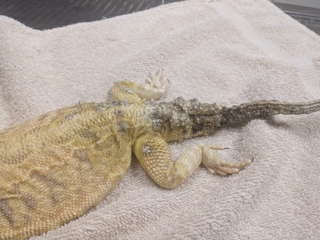
What is “tail rot”? Tail rot is a layperson’s term often used to describe a number of different medical conditions involving the tail and not a true medical term. Actually, this would be like your doctor calling your ear infection “ear rot” or your broken leg “leg rot”. The tail is not “rotting” or decomposing. Instead a number of issues may be causing problems.
The first condition often referred to as “tail rot” is known as Avascular Necrosis. This is when part of the tail dies because the blood supply has been damaged. This results in the tail turning darker in color, getting dry and hard, and (only if far back enough) eventually breaking off.
Damage to the blood supply can be caused by trauma such as being bitten by another animal (or bearded dragon in the same cage), by being shut in a cage door, or other trauma.
The blood supply can also be damaged by the blood vessels being blocked or occluded by what is called an embolus. Emboli, also known as thromboses, are clumps of something such as. blood clots or inflammatory debris that lodge in the blood vessel preventing blood flow. Blood clots can form after a major surgery or with heart disease. Inflammatory or septic emboli happen with deep, chronic infections or in cases of sepsis.
The last and most common reason the blood supply is damaged is retained shed skin causing constriction of the tail and collapsing the blood supply. Usually this only results in damage to the very tip of the tail. This is why proper humidity and hydration is important in pet reptiles. It is also very important to monitor your pet as they shed to make sure all of the shed comes free.
Another condition often referred to as “tail rot” is fungal infections most often caused by Nannizziopsis sp., formerly referred to as “yellow fungus disease” or CANV. Keep in mind, these funguses are not always yellow. These infections start off with crusting of the skin and as they get worse the fungus can invade into deeper tissue. Often these will resemble Avascular Necrosis in advanced stages as the tissue will start to die as well.
Other tail conditions that may end up referred to as “tail rot” include bacterial cellulitis (infection), osteomyelitis (bone infection), or tumors.
How is “tail rot” treated? First an examination by a veterinarian should be performed to determine the actual medical condition your pet has. As we discussed above, “tail rot” is not a medical condition, but a layperson term often used as a catch-all for any problem with the tail. After your veterinarian determines what is happening, they may recommend further testing to look for infection, heart disease, or other conditions. If the damage is too extensive or enough of the tail is affected, surgical amputation under general will be recommended. If the damage is minor or the tail end falls off without surgery, your pet should still be evaluated to determine what caused the damage and if any additional treatments are needed. If there is an infection that need antibiotics for example. If you think your pet may have a problem with their tail, the best thing to do is to schedule an appointment with a qualified reptile veterinarian.
by Heather A. Bjornebo, DVM, DABVP(Reptile-Amphibian Practice), CertAqV
Introduction:
The risk of spreading disease amongst both captive and wild populations continues to rise as tortoises and turtles continue increase in popularity in the pet trade. A number of important contagious diseases have already been identified and can cause significant morbidity and mortality in these species. A better understanding of these illnesses will allow better population management and more prompt initiation of appropriate veterinary intervention leading to improved animal care, survivability, captive breeding, and repopulation efforts. This article will discuss clinical signs of illness, prevention, and the diagnostic modalities available in confirming cause of illness and identifying carriers of contagious disease.
Contagious diseases in chelonians can be broken up within 4 categories: bacterial, viral, internal parasites, and ectoparasites (external parasites).
Bacterial Diseases:
Mycoplasma spp.
Causes: Mycoplasma agassizii & M. testudinum
Species affected:
Terrapene spp.
Gopherus spp.
Testudo spp.
Geochelone spp.
Indotestudo spp.
Pyxis spp.
Clinical signs:
Upper respiratory tract disease
Conjunctivitis
Anorexia
Lethargy
Treatment:
Appropriate prescription antibiotics
Supportive care
Prognosis generally good
Choanal flushes
Treated animals remain lifelong carriers and recurrence of illness common, stress
Pasteurella testudinis
Often associated with respiratory disease
May be found concurrently with Mycoplasma infections
Significance unknown
Affects Gopherusand Geochelone spp.
Chlamydia & Chlamydiophila spp.
Obligate intracellular pathogens
Contagious to a number of vertebrate species
Zoonotic
Clinical signs
Upper and lower respiratory disease
Anorexia, lethargy
Inability to dive
Necrotic foci in the heart, spleen, and liver
Hepatic lipdiosis
Diagnosis – PCR
Treatment – supportive care and appropriate prescription antimicrobial therapy.
Prognosis – fair depending on severity of illness
Salmonella spp.
Appears to be a component of the normal GI flora of reptiles
Zoonotic
Often asymptomatic
Has caused death in isolated cases in captive tortoises and turtles
Suspect opportunistic infection
Clinical signs
Emaciation
Erosions of the plastron
Sloughing scutes
Discolored carapace
Death
Diagnosis – culture
Treatment – supportive care and appropriate prescription antimicrobial therapy
Borrelia & Rickettsia spp.
Borrelia burgdorferi
Zoonotic – Lyme disease
Spread by ticks Ixodes spp. & Hyalommaaeyptium
Isolated from ticks on Testudo spp. in Europe
Borrelia turcica
No human cases documented
Spread by ticks Hyalommaaeyptium
Primary host is Testudo spp.
Rickettsia spp.
Zoonotic – spotted fevers and typhus
Spread by ticks Ixodes spp.
Isolated from ricks on Testudo spp. in Europe
Viral Diseases:
Herpesviruses
At least 12 herpesviruses identified that infect chelonians
Often mistaken for Nutritional Secondary Hyperparathyroidism
Metabolic Bone Disease
Internal Parasites:
Cryptosporidium spp.
Most commonly recognized in snakes and lizards
Being reported with increasing frequency in chelonians
Sonoran desert tortoises
Indian star tortoises
Pancake tortoises
Russian tortoises
Radiated tortoises
Gopher tortoises
Indotestudospp and Testudo spp.
Italy, Germany, Spain, Switzerland, Czech Republic, Ghana, Australia, & US
Suspect transmission similar to that of Cryptosporidium in other host species – Highly contagious!
Clinical Signs
Chronic Diarrhea
Decreased appetite
Pica
Decreased growth rate
Weight loss
Lethargy
Softer than normal shells in juveniles
Passing undigested feed
Diagnosis
PCR with sequencing
Acid Fast examination of fecal smears
Treatment
Currently no data for treatment in chelonians
Paromomycin (Rx) shows promise in some reptile species but all recovered individuals should be considered asymptomatic carriers
Disinfection
Resistant to disinfection
Temperatures greater than 150F (steam or flame thrower)
Formalin (10%)
Glutaraldehyde (2.65%)
Ammonia (5-10%)
Rescue Disinfectant at 1:16 for 15 minutes
Intranuclear Coccidiosis
First identified in 1990
Clinical disease has been described in a number of species
Radiated tortoises
Impressed tortoises
Leopard tortoises
Forsten tortoises
Bowsprit tortoises
Spider tortoises
Galapagos tortoises
Flat-tailed tortoises
Eastern box turtles
Arakan forest turtles
More…
Reported in US and Germany
Most commonly occurs in captive tortoises
Life cycle and route of transmission not fully understood
Morbidity and mortality variable within groups
Clinical signs – non-specific/variable
Anorexia
Lethargy
Lack of normal diurnal behavioral patterns
Increased respiratory effort
Mouth breathing
Rapid weight loss or gain
Mild conjunctival or nasal erythema
Ocular or nasal discharge
Gasping
Subcutaneous edema
Ulceration of the cloacal mucosa
Death can occur within days of clinical signs or after months of treatment
Stress and thermoregulatory changes enhance progression of disease
Reduced immune response
Animals that recover with treatment become asymptomatic carriers
Diagnosis
Identification by cytologic examination of nasal discharge
Biopsy and histologic examination of affected tissues
Quantitative PCR on swabs of the conjunctiva, oral and choanal mucosa, and cloaca
Treatment
Focused on optimal husbandry
Isolation
Supportive care
Hospitalization
Prescription coccidiostats
Prognosis
Guarded to poor as even recovered individuals may have permanent organ damage and succumb months after treatment
Hexamita spp.
Contagious flagellated protozoal parasite that effects the digestive and urinary tracts, including the kidneys, and liver.
Parasite invades kidneys causing damage to the filtration apparatus (glomeruli and renal tubules)
Affected kidneys are enlarged and pale
Leads to kidney failure and death
Documented in cases in numerous freshwater turtle and tortoise species.
Most common cases in practice seen in recently imported Russian Tortosies
Clinical Signs
Failure to thrive
Anorexia
Weight Loss
Death
Treatment
Prescription antiprotozoals
Supportive Care
Environmental decontamination
Cleansing feces off shells to prevent reinfection
Prognosis depends on degree of damage to the kidneys prior to treatment
Nematodes & Trematodes
Nematodes – Hookworms, Roundworms
Trematodes – Flukes
Clinical signs non-specific
Treatment – Prescription antiparasitics
Ectoparasites:
Ticks & Mites
Vectors for other diseases
Anemia with high parasite loads
Treatment
Antiparasitics – not ivermectin!
Physical Removal
Prevention
Disease Prevention:
Establish effective quarantine protocols.
Decontamination and disinfection is very important.
Careful animal health monitoring
Annual veterinary examinations
Fecal parasite screenings
PCR screenings
Conclusion:
Diseases in turtles and tortoises are emerging and popularity of these species are increasing. Understanding the prevalence of these diseases both in natural and captive populations is important in preventing the spread of disease between and within collections.
References:
•Braun, J. et. al. Molecular methods to detect Mycoplasma spp. and Testudinid herpesvirus 2 in desert tortoises (Gpherusagassizii) and implications for disease management. Journal of Wildlife Diseases. 2014. 50(4): 757-766.
•Gibbons, PM. And ZJ Steffes. Emerging Infectious Diseases of Chelonians. Vet Clin Exot Anim. 2013. 303-317.
•Hepner, S. et. al. First investigations on serum resistance and sensitivity of Borreliaturcica. Ticks and Tick-borne Diseases. 2019. 10(5): 1157-1161.
•Heuser, W, et. al. 2014. Soft plastron, soft carapace with skeletal abnormality in juvenile tortoises. Histopathology and isolation of a novel picornavirus from Testudo graeca and Geochelone elegans. Tierärztliche Praxis. Ausgabe K, Kleintiere/Heimtiere. 42. 310-20. 10.1055/s-0038-1623777.
•Kar, S. et. al. Presence of Zoonotic Borreliaburgdorferi sl. and Rickettsia spp. in Ticks from Wild Tortoises and Hedgehogs. Journal of Marmara University Institute of Health Sciences. 2011. 1(3):166-170.
•Origgi, FC. Testudinid Herpesviruses: A Review. Journal of Herpetological Medicine and Surgery. 2012. 22(1):42-54.
•Origgi, FC. et. al. A genomic Approach to Unravel Host-pathogen Interaction in Chelonians: The Example of Testudinid Herpesvirus 3. PLoS ONE. 2015. 10(8):e0134897. doi:10.1371/journal.pone.0134897
•Ng TF, Wellehan JF, Coleman JK, et al. 2015. A tortoise-infecting picornavirus expands the host range of the family Picornaviridae. Arch Virol. 160(5):1319–1323.
•Reavill, DR, and RE Schmidt. 2010. Urinary Tract Diseases of Reptiles. Journal of Exotic Pet Medicine. 19(4): 280-289.
•Rideout, B. Transmissible Infections and Desert Tortoise Translocation: A Comprehensive Disease Risk Analysis. A report of the U.S. Fish and Wildlife Service. June 2015.
•Rosler, R. et. al. Detection of antibodies against Paramyxoviruses in tortoises. J Zoo Wildl Med. 2013. 44(2): 333-339.
•Papp, T. et. al. Paramyxovirus infection in a leopard tortoise (Geochelonepardalisbabcocki) with respiratory disease. Journal of Herpetological Medicine and Surgery. 2010. 20(2-3):64-68.
•Johnson, AJ, et. Al. 2008. RANAVIRUS INFECTION OF FREE-RANGING AND CAPTIVE BOX TURTLES AND TORTOISES IN THE UNITED STATES. Journal of Wildlife Diseases. 44(4):851-863.
•Stilwell, JM, et. al. “EXTENSION OF THE KNOWN HOST RANGE OF INTRANUCLEAR COCCIDIOSIS: INFECTION IN THREE CAPTIVE RED-FOOTED TORTOISES (CHELONOIDIS CARBONARIA),”Joural of Zoo and Wildlife Medicine 48(4), 1165-1171.
Heather A. Bjornebo, DVM, DABVP (Reptile/Amphibian Practice), CertAqV
Introduction
As exotic animal practitioners, we all recognize the important role proper husbandry plays in the health and wellbeing of our patients. Tortoises in general are considered herbivores and veterinarians often fall into a one size fits all trap when it comes to approaching nutrition in this truly diverse group of animals. We must remember that tortoises are as evolutionarily varied as herbivorous mammals. We would not recommend the same diet for a rabbit as we would for an elephant. Can we really recommend the same diet for a Sulcata as we do for a Russian or a Red-foot? So how do we better approach tortoise nutrition as practitioners? With any captive zoologic species this process starts with asking three basic questions: First, what do they eat in the wild? Second, does the captive environment influence their nutritional needs? And third, how do we simulate this in a captive setting?
What are these species consuming in the wild?
Tortoise foraging behavior has been fairly well studied in a number of species commonly kept in captivity. One of the most extensively studied tortoises are the Sonoran desert tortoises of North America, Gopherus morafki. Desert tortoises are opportunistic herbivores foraging 0.5 meters or less off the ground. In general, these tortoises take to desert vines, mallows, grasses, forbs, herbs, and shrubs while occasionally consuming succulents and cacti.1 Leopard tortoises (Geochelone pardalis) have demonstrated similar dietary interest in the wild. A study in Tanzania found that they consumed approximately 74.5% forbs, of which 51% were succulents and 13.5% were legumes. The remainder of their diet was comprised of monocots of which 16.8% were grasses.2
A number of studies involving palearctic tortoises (Testudo sp.) are available within the literature. Fecal samples from 44 Greek tortoises (Testudo graeca) in Algeria were analyzed to evaluate their diet. Plant parts consumed consisted of leaves, stems, and flowers. Their diet consisted of over 70% dicots. Of dicots consumed, the most common were members of Fabaceae, Compositeae, Primulaceae, and Caryophyllaceae families. Monocots were consumed in low numbers and only 3 species were observed in samples: Cynodon dactylon, Hordeum murinum, and Carex remota. No fruit was consumed.3 Two studies involving Hermann’s tortoises (Testudo hermanni) — one in Montenegro and Croatia and the other in Italy — found a preference for legume leaves and to a smaller extent grasses.4,5
While fecal analyses can tell us what is being eaten, they do not provide insight into what foods the tortoises avoided. Two studies have directly observed the animals’ foraging behaviors to answer both of these questions. The first, a study of Testudo graeca graeca in Morocco, found this species to be a rather specialist herbivorous tortoise which focuses its foraging effort under spiny shrubs. The five main plants eaten (70%) were Leontodon saxatilis, Malva parviflora, Astragalus cruciatus, Medicago hispida and Lotus arenarius — all forbs. Eryngium ilicifolium, Emex spinosus, and Spergula flaccida were universally avoided.6 The second is a study of Agrionemys horsfieldi, and while technically they are not considered a member of the Testudo genus, many practitioners still consider Russian tortoises a member of the palearctic group. This study again focused on observing tortoise foraging behavior in the wild and documented both plants eaten as well as those avoided. Their diet consisted of predominantly Ceratocephalus falcatus, Papaveraceae, Koelpinia and Brassicaceae. Grasses were completely avoided by this species despite being abundant.7
Moving back to the Americas, we find another study focusing on Geochelone carbonaria and G. denticulata (red-foot and yellow-foot tortoises, respectively) in Northwestern Brazil. Fecal analysis was utilized to find these tortoises consumed a large variety of fruits, flowers, mushrooms, palm frond bases, unidentified grasses, live leaves, vine stems, roots, insects, and carrion. The ingestion of a large amount of fruits as well as animal matter in these species is in contrast to the species previously discussed. So while these species are still considered generalist herbivores, they do tend to stray to the more omnivorous side of things.8 Another species group also known for their more omnivorous tendencies are the Hingeback tortoises. In a study of Kinixys spekii that focused on food selection in this species, it was noted that wild specimens ingest approximately 47% vascular plants, 41% fungi, and 12% invertebrates (mostly millipedes) in the wild.9
Many other species have been studied, but for the sake of brevity and the fact that these species are less commonly kept in captivity and encountered these shall not be covered in this review. One might note a species that is extremely commonly kept, but studies appear completely absent here. That is Centrochelys sulcata. To date, no English language studies could be located documenting the diet of this species in the wild, which is quite strange given how well known its dietary needs are in the captive environment.
Does the captive environment influence their nutritional needs?
Before we can discuss simulating wild diets in a captive environment, we must acknowledge that being in captivity is different than being in the wild. The question is, how much does captivity impact a tortoise’s nutritional needs? First, let us look at a study on a group of captive Gopherus agassizi (desert tortoises) examining their growth rates over a period of three years. When compared to age matched wild counterparts, their growth rates were markedly accelerated with a one-, two- and three-year-old captive animals being similar in size to 11-, 17-, and 18-year-old wild specimens. This growth rate was attributed to not only skipped winter dormancy for the first two winters but having constant access for high quality nutrition.10 A second desert tortoise study in published in 2018 compared the growth and body condition of tortoises raised outdoors, indoors, and those released directly into the wild. Indoor tortoises grew approximately 16 times faster than those released directly into the wild. Those raised outdoors but fed a captive diet grew 8 times faster than their wild released counterparts. Being raised indoors did have other significant impacts on their health: shell hardness was significantly impacted, and while indoor tortoises were the size of three- or– four-year-olds at only 7.5 months old, they weighed less than those similarly-sized tortoises. It should be noted that indoor raised tortoises were not provided UVB lighting in this study.11 These studies highlight that supplemental feeding can significantly impact growth rates which needs to be a consideration with captive tortoises.10,11
A study in Testudo hermanni found similar results when it compared artificial housing and diets to outdoor housing and naturally growing vegetation. Using dual-energy x-ray absorptiometry, they evaluated bone density and the tortoises’ conformation. Tortoises raised indoors were provided UVB, and no deficits of bone density were noted. However, higher bone density was associated with pyramiding and was noted in both groups housed in artificial conditions.12
Supplemental heating in captivity also needs to be considered. A study in 2002 investigated the effects of supplemental heat on captive Stigmochelys pardalis (leopard tortoises)and Centrochelys sulcata. It found that both growth rate and incidence of shell pyramiding increased with excess nocturnal heat.13 Humidity can also impact growth in captive tortoises. A study in 2002 investigated the influence environmental humidity along with dietary protein impact has on carapacial conformation on Centrochelys sulcata. Drier environmental conditions produced more pyramiding than humid conditions, as did increased dietary protein.14
How do we simulate natural diets in a captive setting?
Although we would ideally feed natural diets to captive chelonians, many diets provided in captivity are based on more readily available grocery store greens, which can also impact tortoise health. There are few studies investigating diet and dietary components in captive tortoises, some including actual feeding trials. One interesting study in Testudo graeca terrestris in Israel investigated diets with the goal to increase growth in a model breeding farm. Vegetarian diets consisting of horticulturally available greens resulted in soft shell syndrome and high mortality in hatchlings. Increasing the protein levels alleviated the soft shell syndrome but resulted in gout and again high mortality. The high protein diet consisted of cat food and corn flour, used to dilute the cat food in an attempt to prevent gout. This diet resulted in sexually mature females by 230 weeks of age and mature males at 170 weeks. However, this study did not discuss any impact on conformation or negative consequences of this rapid growth.15 While this study is definitely informative of the impact protein has on growth rate, the high protein diet is definitely not one we should encourage for appropriate tortoise nutrition in captivity.
In 2005, a study looked into the influence dietary calcium levels have on leopard tortoises. Tortoises where split into four groups. The first group received no calcium supplementation. The second received calcium supplementation based on recommendations by the manufacturer. Groups 3 and 4 received three times and nine times the manufacturer’s recommended doses of calcium. Not surprisingly, tortoises that were not supplemented ended up with depleted calcium levels throughout their bodies by the end of the study. Those receiving the manufacturer’s recommended doses also did not calcify to normal levels. Those with three times the manufacturer’s recommended doses had high growth rate and were thriving; however, both groups 3 and 4 had metastatic calcifications observed postmortem.16
Mineral levels were also the subject of a study on Testudo hermanni. The digestibility of calcium, magnesium, and phosphorus was investigated. Higher calcium concentrations in the feed led to a higher digestibility of not only calcium but magnesium as well. 17
Lickel also investigated diets in leopard tortoises looking at the digestibility of a complete, extruded feed. This commercially available feed was fed to a group of juvenile tortoises for three or seven days per week as part of their diet. Weight, digesta transit time, rate of passage, and indigestible fill were evaluated. Tortoises receiving the diet daily grew more in plastron width and carapace height, but not carapace length. The researcher did not discuss whether this increased carapace height was the result of pyramiding, but her assessment was that those tortoises fed daily did not grow ideally conformationally.18
So… Where does this leave us on understanding the nutritional needs of captive tortoises?
We have come a long way in understanding the dietary needs of these animals. Back in 2005, Innis published recommendations focusing around store bought items such as dandelion greens, cabbage, endive, etc.19 Ten years later we look at recommendations published by Pellet who noted “a fruit, salad and vegetable diet is not ideal for most species.” She recommended Testudo spp. receive various plants and flowers and be provided an edible weed and plant garden. For larger, grass-eating species, she recommended grass grazing and only supplementing them with edible plants and weeds.20 While we still have a long way to go to fully understand the nutritional needs of these fascinating animals, it is important to not lose sight of their wild diets. Horticultural greens fall short in providing for the nutritional needs of these species and the answer to feeding may not be found in the grocery store but instead in the garden center.
References:
Van Devender, TR. 2002. The Sonoran Desert Tortoise: Natural History, Biology, and Conservation. The University of Arizona Press and The Arizona-Sonora Desert Museum; Tucson, Arizona.
Kabigumila, J. 2001. Sighting frequency and food habits of the leopard tortoise, Geochelone pardalis, in Northern Tanzania. African Journal of Ecology; 39:276-285.
Rouag, R.. et al. 2010. Food choice of an Algerian population of the spur‐thighed tortoise, Testudo graeca. African Journal of Herpetology. 57(2), 103-113.
Meek, R. 2010. Nutritional selection in Hermann’s tortoise, Testudo hermanni, in Montenegro and Croatia. B.C.G. Testudo. 7(2):88-95.
Vecchio, et. al. 2011. Seasonal Changes in the Diet of Testudo hermanni hermanni in Central Italy. Herpetologica, 67(3):236-249.
El Mouden, EH et al. 2006. Testudo graeca graeca feeding ecology in an arid and overgrazed zone of Morocco. Journal of Arid Environments; 64:422-435.
Largarde, F., et. al. 2003. Foraging behaviour and diet of an ectothermic herbivore: Testudo horsfieldi. Ecography. 26:236-242.
Moskovits, DK. & KA. Bjorndal. 1990. Diet and Food Preferneces of the Tortoises Geochelone carbonaria and G. denticulate in Northwestern Brazil. Herpetologica. 46(2). 207-218.
Hingeback
Jackson, CG., et. al. 1976. Accelerated Growth Rate and Early Maturity in Gopherus agassizi. Herpetologica. 32:139-145.
Daly, JA., et. al. 2018. Comparing Growth and Body Condition of Indoor-reared, Outdoor-reared, and Direct-released Juvenile Mojave Desert Tortoises. Herpetological Conservation and Biology. 13(3):633-633.
Gramanzini, M. et. al. 2013. Assessment of Dual-energy X-ray Absorptiometry for Use in Evaluating the Effect of Dietary and Environmental Management on Hermmann’s tortoises (Testudo hermanni). AJVR. 24(6).
Heinrich, ML., et. al. 2016. Effect of Supplemental Heat in Captive African Leopard Tortoises (Stigmochelys pardalis) and Spurred Tortoises (Centrochelys sulcata) on Growth Rate and Carapacial Scute Pyramiding. Journal of Exotic Pet Medicine. 25(1):18-25.
Wiesner CS. And C Iben. 2003. Influence of Environmental Humidity and Dietary Protein on Pyramidal Growth of Carapaces in African Spurred Tortoises (Geochelone sulcate). J Anim Physiol a Anim Nutr. (87):66-74.
Lapid, RH. et. al. 2005. Growth and body composition in captive Testudo graeca terrestris fed with a high-energy diet. Applied Herpetology. 2(2):201-209.
Fledelius, B. et.al. 2005. Influence of calcium content of the diet offered to leopard tortoises (Geochelone pardalis). The Veterinary Record.
Liesengang, A. et. al. 2007. Influence of different dietary calcium levels on the digestibility of Ca, Mg, and P in Hermann’s tortoises (Testudo hermanni). Journal of Animal Physiology and Animal Nutrition. 91:459-464.
Lickel, LE. 2010. Intake, apparently digestibility, and digesta passage in leopard tortoises (Geochelone pardalis) fed a complete, extruded feed. A Thesis Presented to the Faculty of California Polytechnic State University San Luis Obispo, CA, USA.
Innis, C. 2005. Considerations in Formulating Captive Tortoise Diets. ARAV. 4(1):8-12.
Pellet, S. et. al. 2015. Tortoise feeding and nutritional requirements. Companion Animal. 21(4):240-245.
by Heather Bjornebo, DVM, DABVP(Reptile-Amphibian Practice), CertAqV
Many clients with rabbits are unaware of the important health benefits of spaying rabbits. Spaying refers to the surgical sterilization of a veterinary patient by removing their ovaries and, at least in the United States, their uterus. There are many benefits to having your rabbit spayed that go beyond preventing unwanted litters.
The biggest benefit is the elimination of the potential to get uterine cancer. Uterine tumors are extremely common in rabbits as they age. Some studies have shown as many as 44% of rabbits developing some form of uterine abnormality in their lifetime. Another important benefit of spaying your rabbit is spayed rabbits are also significantly less likely to get mammary tumors (breast cancer).
Beyond the medical benefits of spaying your rabbit, there are also benefits that will help her make a better pet for your family. Spayed rabbits are better at using their litterbox and tend to be less aggressive. Spay rabbits are easier going and oftentimes more affectionate.
So what should you consider when looking for a veterinarian to have your rabbit spayed? Rabbits in general are considered a higher anesthetic risk than dogs and cats so it is very important to find a veterinary hospital where not only is the veterinarian very familiar with your rabbit’s medical needs, but their staff is trained well so your rabbit receives the best care and monitoring during the procedure. While cheaper prices can be found at spay/neuter only practices, often time this lower cost comes with less one-on-one care, decreased monitoring during and after the procedure, and less supportive care such as skimping on IV fluids. Given the special needs of rabbit patients, we recommend rabbits be spayed at a full service exotics practice. To find an exotic veterinarian in your area visit aemv.org and use their find-a-vet search tool.
By Heather Bjornebo, DVM, DABVP(Reptile-Amphibian Practice), CertAqV
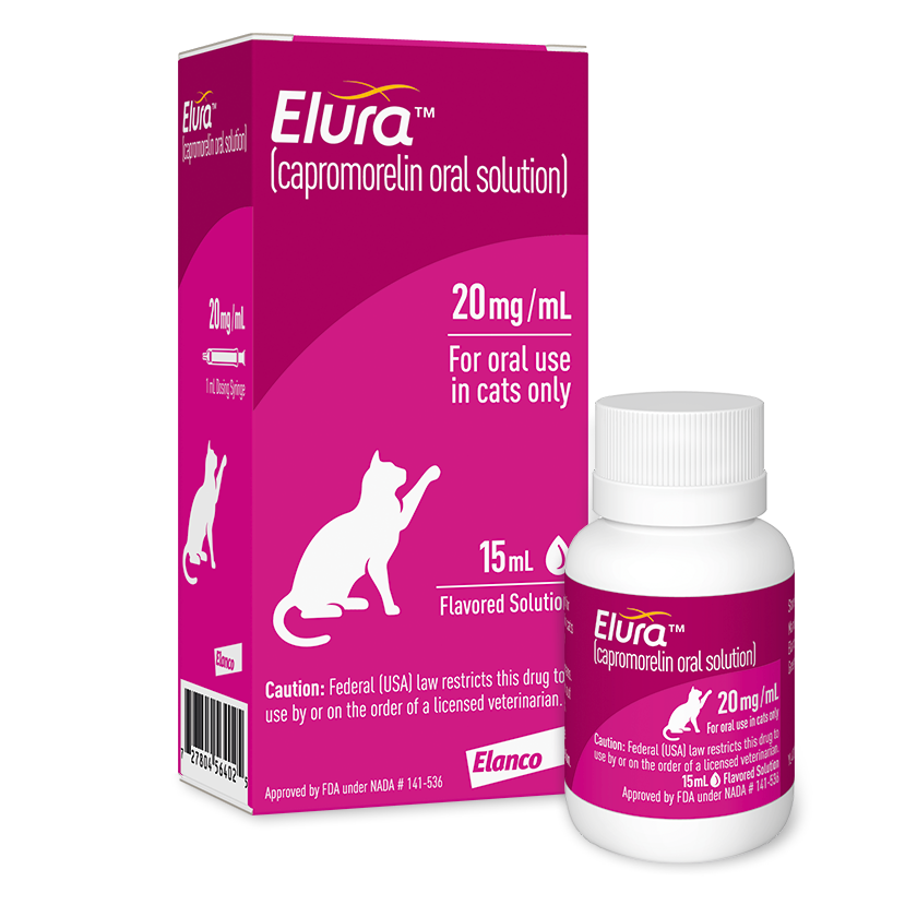
One of the things I love about exotic pet medicine is how rapidly things are advancing and progressing. It seems every year we are getting new advanced treatment options for our patients which is so exciting. One medication that I am excited about is Capromorelin.
Recent studies on capromorelin, sold by the trade names Entyce® (dogs) and Elura® (cats), have shown promise as a medication to help stimulate appetite in sick exotic pets. Classified as a ghrelin agonist mimics the hunger hormone ghrelin where in the brain it acts on the hypothalamus resulting increased growth hormone release by the pituitary gland causing increased hunger and food intake. In addition, capromorelin also increases insulin-like growth factor-1, which increases lean muscle mass.
One recent promising study published in the Journal of Exotic Pet Medicine and Surgery on hedgehogs showed significant increases in food intake when the medication was administered. Seventy percent of the hedgehogs in this study administered this medication showed an increase over those recieving the placebo consuming on average 11% more food.1
A study showed capromorelin decreased stress induced weight loss in laboratory mice. The only study group where no benefit was seen with this medication was when patients were receiving the long acting opioid pain medication Buprenorphine-XR.2
Another recent study in rabbits compared the efficacy of capromorelin versus Mirtazapine on improving appetite. Mirtazapine is another meidcation often used in veterianry medicine to increase appetite in sick animals. It is available as a topical gel that can be applied to the ear lobe. While previous studies in rabbits showed mirtazapine increases fecal output, it did not improve appetite. In this study it showed a positive effect on appetite with healthy rabbits increasing their intake after 2 days. Normal patients given capromorelin also showed significant increase in appetite, though not as profound as those given mirtazapine. Conversely, patients recovering from neuter surgery didn’t demonstrate any significant improvement in appetite on either medication. The overall take home was both medications help appetite, with mirtazapine being more effective and less stressful for the patient due to being topical and not oral. However, both did show promise in treatment of GI stasis.2
Capromorelin has also been studied in birds. In a study in chickens the medication not only biochemically increased their blood insulin levels, it increased their appetite demonstrated by increased pecks per hour.4
Definitely, more studies are needed to fully evaluate the use of this medication in exotic pets. So far the initial studies show a lot of promise, even if the results in rabbits suggest topcial Mirtazapine may be the medication of choice in this species. While less effective, it still gives practitioners another tool to treat GI stasis in this species. Hopefully, in the years to come more studies covering its efficacy and use in many other types of exotic pets, including reptiles.
If you have questions about Capromorelin, contact your family veterinarian.
References:
Huckins, G.L., Mans, C. and Doss, G.A., 2025. Effects of oral capromorelin on food intake and body weight in healthy, four-toed hedgehogs (Atelerix albiventris). Journal of Exotic Pet Medicine, 52, pp.1-3.
Punger, E.M., Norris, S.L., Stevens, S.C., Santos, K.H. and Christy, A.C., 2024. Investigating the Effect of Enterally Administered Capromorelin on Body Weight in Mice (Mus musculus). Comparative Medicine, 74(5), pp.327-335.
Draper, J.M., Savson, D.J., Lavin, E.S., Feldman, E.R., Singh, B., Martin-Flores, M. and Daugherity, E.K., 2022. Comparison of effects of capromorelin and mirtazapine on appetite in New Zealand white rabbits (Oryctolagus cuniculus). Journal of the American Association for Laboratory Animal Science, 61(5), pp.495-505.
Ceron-Romero, N., Taofeek, N., Thomas, A., Vroonland, E., Sanmartin, K., Verghese, M., Heinen, E. and Vizcarra, J.A., 2021. Capromorelin, a ghrelin receptor agonist, increases feed intake and body weight gain in broiler chickens (Gallus gallus domesticus). Poultry science, 100(8), p.101204.
Rabbit hemorrhagic disease (RHDV2) is endemic in southern Arizona, meaning it can’t be eradicated. The disease is highly contagious and can be spread through direct contact with infected rabbits, their feces, urine, and contaminated materials. It can also be transmitted by insects, such as flies, which can carry the virus.
Here are some things you can do to protect your pet rabbit and prevent the spread of RHDV2:
Not using any equipment, clothing, or boots for field work that you use with your pet rabbit
Vaccinate your pet: Get your pet rabbit vaccinated twice as soon as possible. You can get your pet vaccinated at select private exotic animal clinics.
Keep your pet indoors: Ideally, all pet rabbits should be housed indoors.
Take precautions when handling wild rabbits: If you work with or hunt wild rabbits, take special care to prevent your pet rabbit from coming into contact with them. You can do this by:
Removing any field clothing and showering after handling wild rabbits .
Did you know that respiratory infections are come of the most common medical conditions seen in pet rats. Both acute respiratory illness and chronic have been well documented. Acute bacterial pneumonia is characterized by sudden onset of symptoms and is often caused by Corynebacterium kutscheri or Streptococcus pneumonia. Corynebacterium kutscheri is often referred to as pseudotuberculosis (a.k.a. false tuberculosis) and is more often seen in older rats. On the other hand, Streptococcus pneumonia is more commonly diagnosed in younger rats and is actually contagious to people. Chronic disease, commonly referred to as murine respiratory mycoplasmosis, is caused by Mycoplasma pulmonis. Co-infections with cilia-associated respiratory bacillus, Sendai virus, or Sialodacryoadenitis virus are common.
Symptoms of respiratory illness in rats include difficulty breathing, rapid respirations, blue gums, sneezing, ocular/nasal discharge, red pigment staining around the nose, eyes, and front paws, lethargy, head tilt to rolling, muffled heart sounds on veterinary examination, and acute death in severe cases.
Following a thorough physical examination, your veterinarian may recommend chest x-rays. X-rays of the skull may also be recommended to evaluate the inner ear. Blood testing may also be recommended to evaluate the body’s response to infection through a while blood cell count and metabolic state through a serum biochemistry panel. Your veterinarian may also recommend microbial culture and a DNA probe (a.k.a. PCR) test for Mycoplasma especially if the patient isn’t responding as expected to treatment.
Initial therapy involves stabilizing the patient. Hospitalization for oxygen therapy is necessary for patients struggling to breathe. Patients should remain hospitalized until stable on oral medications. Your vet will prescribe appropriate antibiotics and anti-inflammatory medications based on the patient’s illness and needs. Additional treatment with bronchodilators, fluid therapy, and assist feeding. Patients with chronic illness may require long term or pulse therapy as well as may be prescribed medications to treat secondary chronic pulmonary hypertension.
Learn more about respiratory disease in rats by visiting our Educational Guide here.
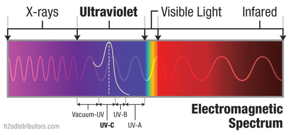
UV light or Ultraviolet light is high energy light than violet but lower energy than X-rays on the electromagnetic spectrum. Ten percent of the light energy produced by the sun is UV light. UV light is invisible to most humans. Small birds, on the other hand, have a forth color receptor in their retinas that gives them “true” UV vision.
UV light is further broken up into UV-A, UV-B, and UV-C.
UV-A is low wave UV. It easily penetrates the ozone layer. This is the light we find in black lights and is known as “soft” UV. Reptiles utilize this light to help regulate behaviors (a.k.a. their circadian rhythm) such as feeding, daytime movement, mating, and similar activities.
UV-B is medium-wave UV or “intermediate” UV and most of this light frequency is absorbed by our ozone layer. Biologically, it is utilized by non-fish vertebrates like reptiles, amphibians, birds, mammals, and even humans for vitamin D synthesis.
UV-C is short-wave UV or “hard” UV. It is ionizing radiation and used in germicidal UV sterilizers. Luckily, the UV-C produced by our sun is completely absorbed by the ozone layer and our atmosphere.
Why do reptiles need UVB light?
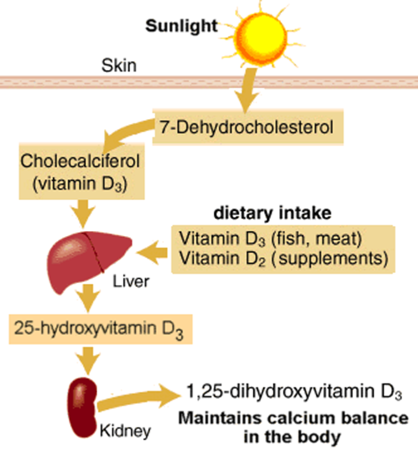
Reptiles, like many vertebrates, utilize UV-B light in the synthesis of vitamin D. When UV-B light interacts with the skin it interacts with 7-dihydrocholesterol activating it into cholecalciferol, or vitamin D3. From there it undergoes further transformation within the liver and kidneys to become 1,25-dihydroxyvitamin D3 used for maintenance of calcium balanced within the body. So without UV-B light that first step in producing this vital chemical regulator within the body never happens. 1,25-dihydroxyvitamin D3 is important as it increases absorption of calcium from the diet, decreases loss of calcium in the urine from the kidneys, prevents overproduction of parathyroid hormone, and modulates the activity if the cells of the skeletal system.
Do all reptiles need UVB light?
Diurnal reptiles, such as iguanas, bearded dragons, turtles, tortoises, water dragons, day geckos, etc. all require UV-B for production of active vitamin D3. Crepuscular reptiles, those active at dawn and dusk, have been found to also be able to utilize UV-B to activate vitamin D3. However, they can also utilize dietary vitamin D3 and have been raised in captivity for generations without UVB lighting. Studies in nocturnal reptiles (i.e. pythons, boas) have shown they cannot utilize UV-B for vitamin D3 activation. However, there may be additional benefits that UV-B lighting provides that have yet to be discovered.
What sources of UVB lighting are available to pet owners?
UV-B light for your reptile can be naturally provided from the sun or artificially by using specially designed light bulbs that produce UV-B. Natural sun can only be used by reptiles housed outside. This is because UV-B cannot penetrate glass so placing your pet near a window will not work. UV-B is only able to penetrate the glass of UV-B bulbs because the glass has been specially treated to allow UV-B to penetrate the glass. This coating wears off over 6-12 months of use causing these bulbs to stop producing UV-B light long before they burn out. This is why manufacturers recommend replacing these bulbs regularly. Artificial UV-B is available in 4 different types of reptile bulbs: fluorescent, mercury vapor, metal halide, and LED.
The first is fluorescent which produces light using a low-pressure mercury-vapor gas-exchange lamp that fluoresces to produce visible light. These bulbs produce minimal heat and come in 3 types. The first are the T8 bulbs, this is a second generation fluorescent straight tube bulb (first generation being T12 bulbs and are no longer produced as UV-B bulbs). The second are the compact fluorescent bulbs where the fluorescent tube is formed into a coil or loop and then set into a normal light bulb socket. Both T8 and compact fluorescent bulbs produce similar amounts UV-B. The third and newest type are the T5 or third generation straight tube fluorescent bulbs. These emit stronger light and have more UV-B production than both T8 and compact fluorescent bulbs.
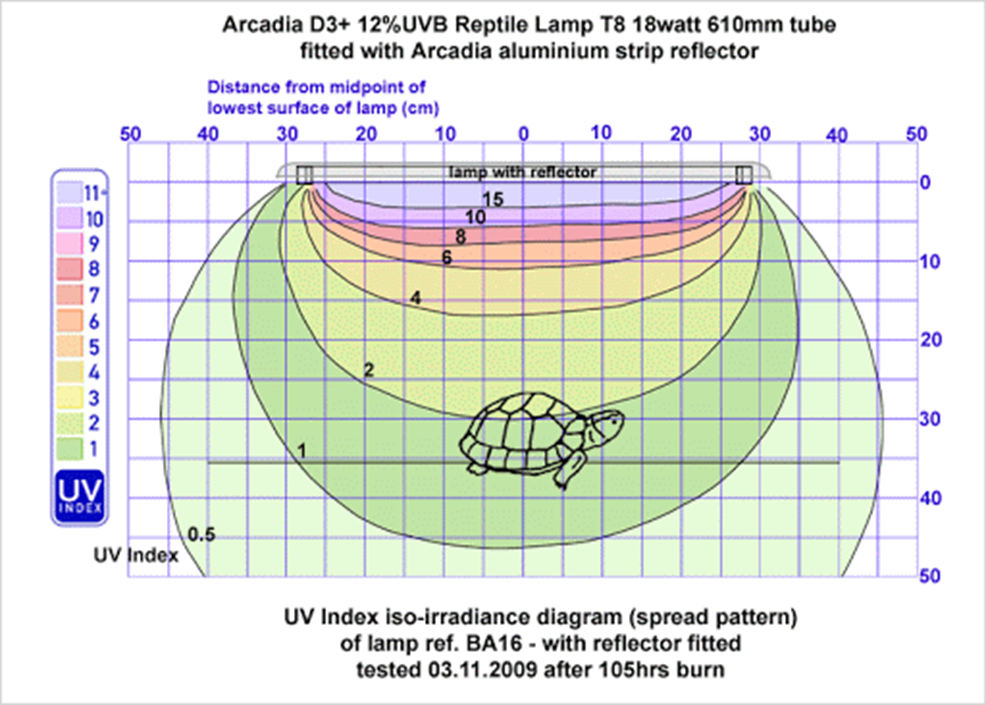
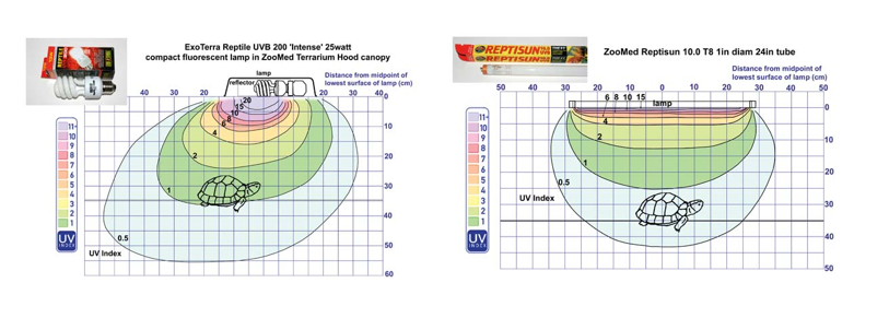
Most UV-B fluorescent bulbs are sold in various strengths, which vary in their labeling depending on the manufacturer. For example, Zoo Med sells them as 2.0, 5.0, and 10.0 bulbs corresponding to 2%, 5% and 10% UV-B production respectively. While Arcadia sells 2%, 7%, 10%, 12% and 14% bulbs. Exo-Terra takes a completely different aproach utilizing a more complex system to help keepers choose the correct bulb based on species and basking distance with bulbs labeled as 100, 150, or 200.
The second type of UV-B bulb available to reptile owners are Mercury Vapor bulbs. A mercury-vapor lamp is a gas-discharge lamp that uses an electric arc through vaporized mercury to produce light. They procude both head and UV-B light. These bulbs are ballasted, when they turn off they need to cool down before they will turn back on, so they cannot be regulated with a thermostat like standard heat lamps. They have a higher UV penetration and UV index than fluorescent bulbs making them ideal for larger enclosures (40 gallons or larger).
Metal Halide bulbs are the third type available on the market. A metal-halide lamp is an electrical lamp that produces light by an electric arc through a gaseous mixture of vaporized mercury and metal halides. This bubl also produces both heat and light, but less heat at a lower wattage than mercury vapor bulbs. These high intensity UV-B bulbs also work well for larger habitats. It should be noted that special fixures are required for these and standard heat domes will not work.
The final type available are LED or light-emitting diode lamps. These are a semiconductor light source that emits light when current flows through it. Relatively new to the market, more research is needed to really gauge how they compare to other bulbs on the market. While expensive, these bulbs are energy efficient and produce little to no heat.
What is UV penetration and why is understanding this important to choosing the right lighting for your pet?
UV penetration is the concept that the farther you get from the UV-B source, the lower the UV-B index is. Different bulbs are going to give you different depths of penetration into the cage. It is important to take this into account when choosing the lighting for your pet. In general, mercury vapor and metal halide bulbs have the highest (or deepest) penetration while fluorescents the penetration is lower. Below are some examples of UV penetration diagrams for a variety of different UV-B bulbs.
What is UV index?
The last area that needs to be touched on is UV index. UV index is an international standard measurement of the strength of sunburn-producing ultraviolet radiation at a particular place and time. It’s an open-ended linear scale, directly proportional to the intensity of UV radiation that causes sunburn on human skin. It was invented to help people effectively protect themselves from UV radiation, which has health benefits in moderation but in excess causes negative effects. We borrow this in reptiles just as a measure of the amount of UVB reaching our pets.
Summary
UV-B lighting is essential for the health of many pet reptiles. Most reptiles require UVB lighting to properly absorb and utilize dietary calcium. Without UVB many reptiles will develop Nutritional Secondary Hyperparathyroidism a.k.a. “Metabolic Bone Disease.” It is important to understand that artificial UVB lights need to be changed every 6-12 months as they will stop giving off UVB light. Both species and cage setup should be considered when choosing the right UVB source.
Reptiles are often referred to being “cold-blooded”, which can be misleading. More appropriately they should be considered poikilothermic or ectothermic, which means that unlike mammals and birds reptiles are unable to regulate their body temperatures internally and change their body temperature in adaptation to their environmental temperature. Because reptiles do not need to expend as much energy heating their bodies, they have a much lower metabolic rate than that of mammals. Each reptile species has what is referred to as its preferred optimal temperature zone which is a narrow temperature range at which they are active and undergo typical functions such as feeding, digestion and reproduction. Outside of this range these functions may be hindered or cease altogether. Some species will hibernate during colder months and during this time their metabolic rate will decrease.
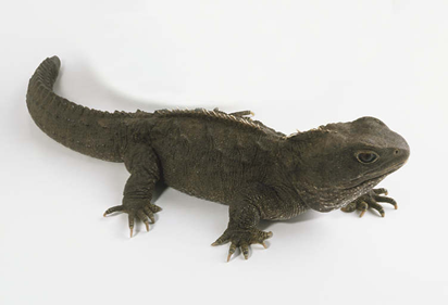
Reptiles belong to the Class Reptilia, which can be broken further down into 4 Orders: Squamata, Chelonia, Crocodilia, and Tuatara.
Tuatara
The Tuatara is an ancient type of reptile only found in New Zealand and is a protected species only rarely seen in captivity in zoos or very special private collections.
Crocodilia
Crocodilians include crocodiles, alligators, and caiman. Members of Crocodilia are illegal to own as pets in Arizona.
Squamata
The order squamata can be broken down into 2 suborders: Serpentes (snakes) and Sauria (lizards).
There are over 2,700 species of snakes worldwide and snakes can be found on every continent besides Antarctica . While the obvious answer to how snakes differ from lizards might appear to be that they lack limbs, it gets confusing when you consider legless lizards. Thus, to be more precise, snakes differ from lizards by lacking external ears and lacking remnants of forelimbs. That said, some snakes do have vestigial hind limbs, which can be seen externally as a pair of spurs on either side of their cloaca. Snakes also lack eyelids. Instead their eyes are protected by a clear scale known as a spectacle, which is shed at the same time as the snake sheds its skin.
All snakes are carnivorous, most feed on rodents, lizards, birds, and even eggs. Since snakes are unable to chew or take bites of their meals, they must swallow their food whole. To do this they have a very flexible skull and jawbones. Snakes can easily consume an animal as big as they are wide. The majority of snakes are non-venomous and kill their prey by constriction. Those that are venomous produce venom in glands located behind the eyes and inject it via fangs by biting. Venomous snakes should be discouraged as pets due to the risk of injury to humans.
Most snakes lay eggs (oviparous), with the exceptions of nearly all boas and some vipers such as rattlesnakes who give live birth (viviparous). Snake eggs are oval shaped and have flexible, leathery shells.
Snakes are largely non-social and live solitary lives, rarely seeking out company outside of the breeding season.
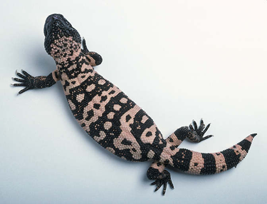
Lizards far outnumber snakes with nearly 5,000 different species worldwide. And like snakes, lizards can be found on every continent except Antarctica. Lizards have well developed color vision and communicate via body language and pheromones. Most lizards are solitary animals and keep a loose territory. Most lay eggs (oviparous) but some do give live birth (viviparous). There are two species of venomous lizard, the Gila monster and the beaded lizard.
Some lizards have unusual adaptations. Many species of gecko have adhesive lamina on their toes to enable them to climb on sheer surfaces. Many geckos also lack eyelids and, similar to snakes, have a spectacle.
Many species of lizard have the ability to drop their tails, an adaptation referred to as autonomy. Most lizards will regrow their tails with the exception of Rhacodactylus geckos. Lizards that do not practice autonomy will not regrow a lost tail, however.
Both snakes and lizards shed their skin as they grow. Snakes shed their skin in one large piece, while lizards often do so in multiple smaller pieces. Proper shedding is very important to their health.
Chelonians
This suborder includes turtles, terrapins and tortoises. Turtles most accurately should be used to describe sea turtles, which are not kept as pets due to their size and care requirements. Terrapins are the freshwater and land dwelling species that are what we commonly refer to as turtles. Terrapins are omnivorous to carnivorous. Tortoises are strictly land dwelling and predominantly herbivorous. Often, the confusion arises when trying to distinguish a box turtle from a tortoise. While they are primarily land animals, box turtles are terrapins. They are omnivores and have limbs more similar to that of a terrapin.
All Chelonians have a protective shell, however some may be more protective than others. Soft shelled turtles, for instance, have a shell covered with leathery skin that does very little for protection. Other than soft shells, a chelonian’s shell is comprised of living bone covered with plates of keratin, similar to scales, called scutes. It is normal for terrapins to shed their scutes as they grow, but not for tortoises.
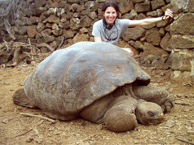
One of the most well known characteristics of chelonians is that they are very long lived; even the shortest-lived aquatic terrapins routinely live 20-30 years. Tortoises can live 60-100 years, depending on species. Chelonians are very social animals and establish loose territories. Males can be very territorial and will fight over resources and females. All chelonians lay eggs (oviparous); they produce eggs with a hard shell similar to a bird’s egg and are usually spherical.
Basic Anatomy
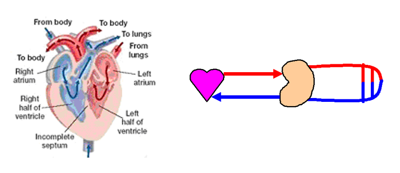
Most reptiles have a 3-chambered heart as well as a renal portal system where blood passes through the kidneys both as it leaves the heart and as it returns to the heart after traveling through the back end of the body.
Reptile lungs are simple and sac-like and often have a vascular epithelium towards the front and non-respiratory epithelium towards the back. They do not have aleoli like mammals, but instead their pulmonary functional unit is called faveoli, small air spaces in the lungs that resemble honeycomb cells.
Reptile gonads are internal and the right and left gonads are located at different levels of the snake between the pancreas and the kidneys. Male snakes and lizards have paired copulatory organs called hemipenes while male chelonians and crocodilians have a single phallus. These are not associated with the urination and only function for mating, thus if diseased these may be amputated.
Snakes
Snakes possess very elastic to allow for ingestion of prey. Periodically shed whole, usually between 2 to 4 times per year depending on growth rate. They also are very delicately built compared to other reptiles. They have two rows of teeth on the top (maxillary and pterygoids) and one on the bottom (mandibular). Their teeth are recurved and are periodically shed throughout life. Their tongue is long, deeply forked and lies in sheath under the glottis and is used in olfaction (smell) together with the Jacobson’s organ found on the palate. They have no external ears and no tympanic membrane or middle ear, yet they are able to hear low frequency sound using the inner ear. Their vision geared for movement and they have no eyelids, instead their eyes are covered with a spectacle. Heat-pits are present on the heads of boas, pythons, and pit vipers allowing them to find prey in complete darkness.
The right lung is larger than the left in snakes, with the left lung completely non-functional (vestigial) in colubrids like corn snakes and kingsnakes. Like all reptiles, snakes lack of diaphragm.
Venom glands in snakes are modified salivary glands and located behind the eyes on the head.
Like all reptiles, the snake ureter empties into part of the cloaca called the urodeum. Snakes lack a urinary bladder. Male snakes have a sexual portion of the kidney that enlarges during the breeding season and produces seminal fluid.
Lizards
Unlike snakes, most lizards shed their skin in pieces. Their skin is usually fairly thick and scaled, with the exception of geckos who possess very thin skin. Many practice tail autonomy, meaning they can drop their tails as a defence mechanism. While most species will regrow their tails, not all can do this, i.e. crested geckos. Bearded dragons do not drop their tails and their tails cannot regenerate if lost.
Their ear drum, or tympanic membrane, is visible and there is no external or ear lobe.
Most lizards have eyelids, with the exception of some species of geckos, and have well developed color vision. Many also possess a degenerate 3rd eye or parietal eye located on the top of the head. It contains a retina and lens and is used to stimulate hormone production as well as moderate behavior associated with thermoregulation and basking times.
The heart is 3-chambered and located between the shoulders in most lizards, with the exception of monitor lizards where it is located low in the chest.
Lizard tongue morphology varies based on function.
Chelonians
Probably the most promanent part of chelonian anatomy is their shell witha dorsal carapace and ventral plastron. Their pectoral and pelvic girdles are uniquely located within their rib cage. Comprised of living bone covered with protectie keratin, their shells are well innervated and can feel touch and pain.
Their eyes are similar to birds and they possess scleral ossicles, small circular bones embedded in the whites of the eyes.
The chelonian ear is hidden behind a large scale or several smaller scales that overlies their tympanic membrane (ear drum).
Chelonians have large lungs that extend the length of the shell. Like all reptiles, they lack a diaphragm. Their shell prevents movement of the rib cage so respiration requires movement of the head and forelimbs.
While chelonians have a relatively simple stomach, the large intestines of many tortoises is extensive and used for microbial digestion of fiber.
Measures all bivalent cations, majority is dissolved magnesium and calcium
Measured in “dH” aka degree hardness— 1 dH=17.8ppm CaCO3
Hardness of the water plays into the buffering ability of the water
Hard water >300 mg/L, soft water <150 mg/L
Used for skeletal and scale growth, and in mollusks and crustaceans for shells
High levels can affect drugs administered in the water and require higher concentrations of drug be added
Too high: zinc deficiency cataracts due to competition for absorption with calcium, nephrocalcinosis
Too low—decreased buffering capacity causing acid pH in environment (see KH and pH information on each parameter), slow or stunted growth (due to not enough calcium for growth)
Carbonate Hardness (KH)
Should be > 4.5 dH (degree of hardness), 7-9.8 dH in coral or invertebrate tanks
Other terms used “buffering capacity”, “alkalinity”
Measure of the carbonates and bicarbonates within the system
Carbonates provide buffering capacity and an energy source for use by the bacteria involved with biofiltration and the plants in the tank when CO2 levels fall
The higher the KH, the more buffering capacity of the water, the larger ability to bind H+ ions and resist pH drop in the water
Important for corals and invertebrate skeleton
If the buffering capacity is used up and not replaced or Hardness level <4.5 dH the everyday processes that occur in the tank with continue to drop the pH = biofilter will stop working, fish will become stress and undergo changes due to acidic irritation
Treatment: Water changes with help replace the buffer, Limestone or baking soda can be added to provide buffer
Phosphate
Phosphate levels <0.1 mg/L
In high levels will cause algal over growth
Naturally occur due to breakdown of plants, animals, waste products, and fertilizers
High levels can occur at the water source or from nutrient build up and breakdown in the water (over stocking, algae die-off, lack of water changes, fertilizer application nearby)
Treatment: Water change to decrease, clean the tank/pond of extra plant or algae and debris that you can. if large scale you can place a phosphate binding filter or phosphate binding chemicals into the water.
Copper
Aim for zero ppm, no more than 2.0 mg/L during copper baths
Higher in soft water
Cause- high levels from water source, treatment with bath, algae treatment
Toxicity: osmoregulation dysfunction causing high levels of blood potassium from kidney failure/toxicity, immunosuppression, damage to lateral line organ, generalized multi-organ